description
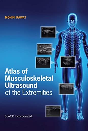
0Featuring nearly 700 illustrations, images, and photos, Atlas of Musculoskeletal Ultrasound of the Extremities by Dr. Mohini Rawat is a comprehensive visual guide to musculoskeletal ultrasound imaging for health care students and clinicians. Musculoskeletal ultrasound imaging is a new, rapidly growing field with applications across many health care disciplines. With its increased popularity comes a need for detailed training resources. The Atlas of Musculoskeletal Ultrasound of the Extremities presents information on scanning protocols for the joint regions and peripheral nerves of the upper and lower extremities in an easy-to-follow, highly visual format. Beginning with an overview of ultrasound physics, equipment, terminology, and technique, the book provides detailed instruction for musculoskeletal ultrasound of the shoulder, elbow, wrist, hip, knee, ankle and foot, concluding with a comprehensive chapter on peripheral nerves. Each chapter contains detailed images of scanning protocols, anatomy, sonoanatomy, patient positioning, and probe positioning for each joint region. Images are accompanied by explanatory text descriptions, along with clinical pearls under points to remember. Designed for students and clinicians in physical therapy, occupational therapy, athletic training, orthopedics, rheumatology, physiatry and podiatry, the Atlas of Musculoskeletal Ultrasound of the Extremities provides essential introductory training materials and serves as a helpful reference for busy clinical environments.
member goods
No member items were found under this heading.
listens & views

MOB ACTION AGAINST THE STATE ...
by MOB ACTION AGAINST THE STATE / VARIOUS
COMPACT DISCout of stock
$18.25
Return Policy
All sales are final
Shipping
No special shipping considerations available.
Shipping fees determined at checkout.


